Services
Diagnostics
If you’re concerned your horse or pony is out of sorts, you can book a consultation with equine vet Carlos to investigate the problem.
Equine diagnostics uses advanced methods like imaging and blood tests to identify horse health issues accurately. These tools are crucial for tailored treatment, ensuring equine well-being and performance. They enable veterinarians to provide optimal care for their patients.
Endoscopy
Upper Airway Endoscopy is carried out using our flexible 1.3m fibro endoscope. It can be used to diagnose upper and lower airway problems such as laryngeal hemiplegia, dorsally displaced soft palates, guttural pouch problems or infections. Samples can be taken to evaluate the cells in the respiratory tract for diagnosis. Urethral endoscopy can also be performed in the yard. For other procedures such as arthroscopy, laparoscopy horses are referred to a local specialist centre. Gastroscopy (visualization of stomach for ulcers) could be arranged to be performed at the yard.
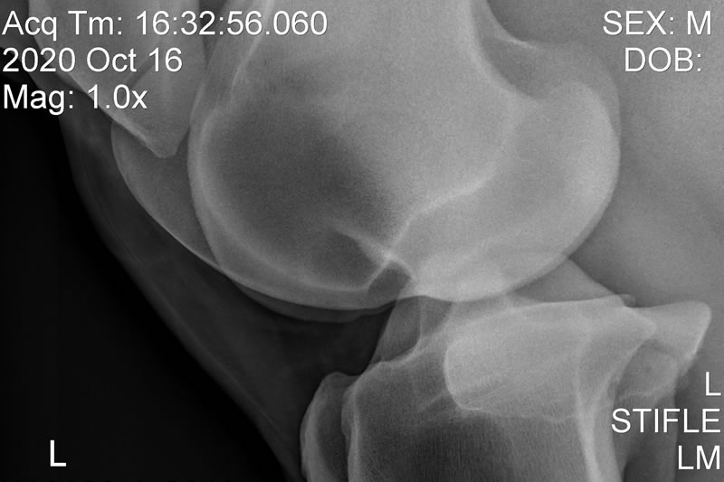
Radiology
Taking X-rays at the yard with our fully portable X-ray unit allows us to diagnose and manage limb conditions in the comfort of the stable, liaising and sharing images immediately with potential purchasers, farriers and referral centres.
Echography / Ultrasound Scanner
With some injuries, especially tendon / soft tissue injuries, we will recommend you have an ultrasound scan of the area as this is invaluable in determining which structure is damaged and its severity. Ultrasound scans are generally done 7-10 days after the initial injury and repeated at intervals of 3-4 months to monitor progress. All this is done in the comfort of your stable.
Electrocardiography (ECG)
At the yard we take a measurement of your horse’s electrical activity by connecting our ECG machine to specific points on the horse using electrodes. A “trace” is produced that shows the net movement of electrical current at any given time.
The signals are shown as waves which represent specific events in the cardiac cycle.
- P Wave – represents atrial depolarisation and contract
- QRS Complex – represents ventricular depolarisation and contract
- T Wave – represents both atrial and ventricular depolarisation
Together with heart rate, heart rhythm, audible murmurs, timings measurement – for example the P-R interval, the R-R interval and the S-T interval.
Rhythm is assessed on the ECG trace too, making sure that every P wave is followed by a QRS Complex.
Clinical Examinations
Careful, detailed and thorough clinical examinations are the most commonly used diagnostic tool of all, with the aid of a thermometer, and ophthalmoscope, stethoscope, a pen torch, hands, eyes and careful questioning.